Latest Entries »

Sleep Related Hypoventilation-Hypoxemic Syndromes:

Sleep Related Non-obstructive Alveolar Hypoventilation (Idiopathic):

  • Diagnostic Criteria
    • PSG demonstrates shallow breathing for longer than 10 seconds with associated oxygen desaturation, frequent arousals or brady-tachycardia
    • No primary lung diseases, skeletal abnormalities or neuromuscular disorder
    • Elevated PCO2 at night but may eventually persist into the day
  • Distinguished from CSA/OSA by sustained hypoxemia instead of brief and cyclical (often last several minutes)

Congenital Central Alveolar Hypoventilation Syndrome:

  • Diagnostic Criteria
    • Infant exhibits shallow breathing, or cyanosis or apnea during sleep. (In severely affected infants pulmonary HTN and cor pulmonale may be evident)Hypoventilation worse in sleep as compared to wake
    • The rebreathing response to hypoxia or hypercapnia is absent/diminishe
    • PSG shows severe hypercapnia dn hypoxia predominantly without apnea
  • The majority of cases have been found to occur in association with mutations of a PHOX2B gene.

Blood Gases

The normal PO2 in young adults averages 95mmHg (range 85-100mmHg).

Note that above a PO2 of 60mmHg the curve is fairly flat and cyanosis is probably undetectable.

There are five causes of Hypoxemia:

  1. Hypoventilation
  2. Diffusion impairment
  3. Shunt
  4. Ventilation-Perfusion inequality
  5. Reduction of inspired PO2.

There are two major causes of increased arterial PCO2:

  1. Hypoventilation
  2. Ventilation-Perfusion inequality

Acid – Base Changes

General Rule: For every 10mmg increase in PCO2 the HCO3 will increase by 3-4 chronically and 1 meq/L acutely. The pH will change 0.08 chronically and 0.03 acutely.

Pulmonary Function Tests


  Normal Range
FEV1 80-120% predicted
FEV1/FVC ≥ 0.70
TLC 80-120% predicted
Diffusing Capacity >75-80% predicted


Patterns of Spirometry

  FEV1/FVC FEV1 FVC TLC
Obstructive Decreased Decreased Normal/Decreased Normal/High
Restrictive Normal/High Decreased Decreased Low

 Ventilation in Obesity

  Simple Obesity OHS
CO2 production Increased Increased
Tidal Volume Normal Reduced 25%
Respiratory Rate Increased by 40% Increased by 25%
Minute Ventilation High Low
Vd/Vt Normal High
PCO2 Normal Increased

 With simple obesity the ERV (expiratory reserve volume) can be decreased. With obesity hypoventilation syndrome ERV, RV, and TLC are decreased.

Obesity hypoventilation syndrome is defined as the combination of obesity (body mass index above 30 kg/m2), hypoxia during sleep, and hypercapnia during the day, resulting from hypoventilation. In contrast to patients with primary central sleep apnea who have a high ventilatory response to CO2, patients with OHS have a reduced ventilatory response to PCO2. Therefore, patients with primary central sleep apnea are hypocapnic, whereas patients with OHS are hypercapnic.

Sleep’s effect on Ventilation:

  • During NREM and REM sleep (as compared to Wake) tidal volumes decrease while respiratory rate remains the same.
  • Ventilatory response to hypercapnea is Wake>NREM 3> NREM 2>REM.


 Central Sleep Apnea Syndromes

Primary Central Sleep Apnea: idiopathic disorder which is typically seen in patients with low normal arterial PCO2 (< 40mmHg). Patients present with sleep fragmentation, EDS, or insomnia. A high ventilatory response to CO2 seems to be a predisposing factor. (An increased hypercapnic ventilatory response). These patients have low daytime and sleeping PCO2.  Insomnia and nasal obstruction are thought to potentially exacerbate the condition. There is little evidence that the disorder leads to any cardiovascular consequence.

  • PSG shows: ≥ 5 central apneas/hr. The apnea cycle typically is < 45 seconds.

Cheyne Stokes Breathing: Cheyne Stokes breathing is characterized by recurrent apneas, hypopneas, or both alternating with prolonged hyperpneas during which tidal volume waxes and wanes in a crescendo-decrescendo fashion. The prevalence in patients with CHF is 25-40%. The apnea cycle typically is > 45 seconds (usually 60-120 seconds)

  • PSG shows: ≥ 3 cycles of crescendo-decrescendo breathing and at least one of the following: ≥ 5 central apneas per hour or cyclic crescendo-decrescendo breathing for ≥ 10 consecutive minutes. The

High Altitude Periodic Breathing: Diagnostic criteria: recent ascent to an altitude of ≥ 4000 meters. The condition is believed to be a product of hyperventilation induced by hypoxia encountered at altitude, which leads to hypocapnic alkalosis. Because PCO@ is the principal stimulus to respiration during NREM sleep, a low PCO2 can yield a depressed respiratory drive.

  • PSG shows: recurrent central apneas at a frequency of > 5 per hour. The apnea cycle is typically 15-30 seconds.

Central Sleep Apnea Due to Drug: A variety of abnormalities have been described including central apneas, Biot’s breathing pattern, and periodic breathing. (Biot’s breathing pattern is characterized by groups of quick, shallow inspirations followed by regular or irregular periods of apnea)

  • PSG shows: recurrent central apneas at a frequency of > 5 per hour, periodic or Biot’s breathing pattern

Primary Sleep Apnea of Infancy: Characterized by prolonged central, mixed, or obstructive apneas or hypopneas associated with physiological compromise (hypoxemia, bradycardia, or need for stimulation or resuscitation). It can be secondary to immaturity of the brainstem or secondary to medical conditions that produce apnea by depressing central respiratory control. The frequency of respiratory events increases during REM sleep. Paradoxical chest wall movements are common during active sleep in infants and may cause a fall in PO2 due to V/P defects associated with a decrease in FRC. The prevalence varies inversely with age. 25% of infants with birth weight < 2500g and 84% of infants with birth weight < 1000g.

  • Apnea of Prematurity: Prolonged central apneas lasting ≥ 20 seconds (or shorter duration events that included obstructive or mixed respiratory patterns and are associated with a physiological compromise) are recorded in an infant < 37 weeks.
  • Apnea of Infancy: Prolonged central apneas lasting ≥ 20 seconds (or shorter duration events that included obstructive or mixed respiratory patterns and are associated with a physiological compromise) are recorded in an infant < 37 weeks.  

Obesity hypoventilation syndrome is defined as the combination of obesity (body mass index above 30 kg/m2), hypoxia during sleep, and hypercapnia during the day, resulting from hypoventilation. In contrast to patients with primary central sleep apnea who have a high ventilatory response to CO2, patients with OHS have a reduced ventilatory response to PCO2. Therefore, patients with primary central sleep apnea are hypocapnic, whereas patients with OHS are hypercapnic.

Pediatric Sleep Development

  1. Trace Discontinue – appears at 26-30 weeks
  2. Delta Brushes – appear at 28-30 weeks, disappear at term
  3. Trace Alternant – appears at 32-36 weeks, disappears at 12 weeks post-term
  4. Rapid Eye Movements – appear at 30 weeks
  5. Spindles – 2 months
  6. K complexes and Slow Wave Activity – 4 to 6 months
  7. Ability to discriminate N 1/2/3 sleep stages – 5 to 6 months
  8. Vertex waves – usually first appear at 16 months
  9. Dominant Posterior Rhythm
  • @ 2 months – 4Hz
  • @ 6 months – 6Hz
  • @ 3 years – 8Hz
  • @ 9 years – 9Hz
  1. Posterior slow waves of youth – intermittent runs of bilateral, often asymmetric 2.5 – 4.5HZslow waves superimposed on the DPR, most common in 8 -14 year-olds.
    1. Hypnagogic Hypersynchrony (HH) – Paroxysmal bursts of diffuse high amplitude sinusoidal 75-350µV, 3-4.5Hz waves which begin abruptly often maximal over the frontocentral regions. HH often disappears with deeper stages of NREM sleep. HH is seen in 30% at 3 months, 95% of children between 6-8 months, and is less prevalent after age 4-5.
    2. Sleep architecture in infants:
      1. Term: 50% REM/50% NREM   
      2. 6-12 months: REM 30-35%, NREM 65-70%
      3. 3-5 years: REM 25%, NREM 75%
      4. Sleep duration:
        1. Newborn: 16 hours.
        2. 3 months: 5 – 6 hour major sleep period at night
        3. 5 years: major overnight period of sleep and one nap

Stage N sleep:

  1. No recognizable spindles or K complexes
  2. < 20% high amplitude 0.5-2Hz activity

Stage R sleep: REM lasts 50-60 minutes with up to 10 cycles per major sleep episode

  1. Low voltage, mixed frequency
    1. 7 weeks post-term: 3Hz
    2. 5 months post-term: 4-5Hz  
    3. 9 months: 4-6Hz

Restless Legs Syndrome: Defined by 4 cardinal symptoms as can be recalled by the acronym, URGE:

  • U= urge to move the legs, usually associated with unpleasant leg sensations
  • R= ret induces symptoms
  • G= getting active brings relief
  • E= evening makes symptoms worse

Pediatric Diagnosis: The child meets the above 4 criteria, but, cannot relate a description consistent with leg discomfort, but displays at least TWO of the following three findings:

  • A sleep disturbance for age
  • Parent/sibling with RLS
  • PSG with PLMI of ≥ 5

Demographics: 5-10% of Northern European population. 1.5 x more common in women.

Predisposing factors: Primary RLS occurs independently of other disorders, but may be exacerbated by other factors, whereas secondary RLS is precipitated by other disorders and resolves when they are treated. Pregnancy, Iron deficiency, peripheral neuropathy, radiculopathy, Parkinson’s disease, spinocerebellar atrophy, MS, Charcot Marie Tooth, and renal insufficiency can precipitate RLS. Several medications can aggravate RLS as well including: antihistamines, dopamine receptor antagonists, and most anti-depressants, with the exception of bupropion with its dopamine promoting activity.

Familial Patterns: > 50% report family history. The risk is 3-6 times greater among those with first-degree relatives.

PSG findings: 80% have PLDMS.

PLMS (Periodic Limb Movements of Sleep) – occur in 30% of adults over 50 years of age and in 80% of patients with RLS. To diagnose PLMS one needs to identify ≥ 4 LMs separated by a period between 5 – 90 seconds. See scoring rules below:

PLMS scoring rules

  • ≥ 4 LMs separated by a period between 5 – 90 seconds.
  • Leg movements on 2 different legs separated by < 5 seconds count as a single leg movement.
  • LMs occurring 0.5 seconds before/after an anpea or hypopnea are not to be scored.
  • PLM is to be considered associated with arousal if the arousal occurs within 0.5 seconds.
  • Impedence should be < 10,000Ω although < 5,000Ω is preferred.
  • Leg Movement: 0.5 – 10 second duration, amplitude ≥ 8µV above resting EMG. The end of a LM is defined as a period lasting ≥ 0.5 seconds during which the EMG does not exceed 2 µV above resting EMG

Conditions associated with PLMS: RLS, Narcolepsy, RBD, Huntington’s Disease, Parkinson’s disease, Tourtette’s Syndrome, Stiff Person syndrome, Motor neuron disease, peripheral neuropathy, ESRD, & pregnancy.

Treatment: Treat underlying condition if possible, Dopamine agonists (most common side effect is nausea), neurontin, and opiates.  

Rhythmic Movement Disorder: repetitive, stereotyped movements occurring in drowsiness and sleep, typically seen in infants and children. At 9 months of age, 60% of infants exhibit some form of sleep related rhythmic movement. By 5 years of age only 5% exhibit the behavior. To be classified as a disorder the behavior must result in a significant complaint manifested by one of the following: interference with normal sleep, impairment in daytime function, or bodily injury.

Benign Sleep Myoclonus of Infancy: Repetitive myoclonic jerks of the whole body, trunk or limbs, typically evident between birth and 6 months of age, only during sleep. The movements stop abruptly and consistently when the infant is aroused. These should be differentiated from myoclonic seizures and hyperexplexia, drug withdrawal, and sleep starts.

Hypnagogic Foot Tremor & Alternating Leg Muscle Activation: HFT is a rhythmic movement of the feet or toes occurring at the transition of wake and sleep or during NREM (stages 1 & 2) sleep. ALAM consists of brief activations of the anterior tibialis in on leg in alternation with similar activation in the contra-lateral leg during sleep or arousals from sleep. The significance of these movements is unknown.

Diagnostic Criteria for HFT:

  1. Burst potentials last 0.25 – 1s and occur at a frequency of 0.5-3Hz.
  2. Trains last 10 or more seconds

Diagnostic Criteria for ALMA

  1. PSG shows ≥ 4 ALMA with < 2 seconds between activations.
  2. EMG activations last 0.1 – 0.5s and occur at a frequency of 0.5-3Hz.
  3. Sequences last between 1-30 seconds and may recur

Excessive Fragmentary Myoclonus: PSG shows brief 75-150 msec potentials in various muscle groups occurring asynchronously and asymmetrically with more than 5 potentials per minutes for at least 20 minutes of consecutive NREM sleep.

Insomnia

Insomnia – a complaint of a difficulty initiating or maintaining sleep or reporting non-restorative sleep despite adequate opportunity for sleep, which results in some form of daytime impairment. Overall prevalence of chronic insomnia 10-15%, increasing to 35-50% in persons older than 65. Risk factors include: female sex, elderly, unmarried, unemployed, and those with medical or psychiatric conditions.

  • Patients with chronic insomnia have a 30-50% prevalence of psychiatric disorder, most often depression.
  • Health Risks: Chronic insomnia is associated with an increased rate of medical and psychiatric illness, traffic accidents and reduced QOL.

Adjustment Insomnia – defined as an acute sleep disturbance, which lasts < 3 months and is temporally associated with an identifiable stressor. The sleep disturbance is expected to resolve when the stressor resolves or the subject adapts.

Psychophysiological Insomnia – Insomnia for ≥ 1 month with evidence of conditioned sleep difficulty as indicated by one or more of the following: excessive focus on sleep, ability to sleep better away from home, heightened somatic tension, etc. MSLTs typically reveal heightened alertness, with latencies in the 10-15 min range.

Paradoxical Insomnia – Insomnia for ≥ 1 month owing to sleep state misperception.

Idiopathic Insomnia – The course is chronic as indicated by onset during infancy/childhood, no identifiable cause, persistent course without remissions.

Familial Fatal Insomnia – Rare disorder linked to a mutation of the prion protein gene on chromosome 20. Onset 35-60.

Cognitive Behavioral Therapy – Stimulus Control, Sleep Restriction, Relaxation training, cognitive therapy, and sleep hygiene education.

  • Stimulus Control – don’t spend time in the bedroom when not sleeping
  • Sleep Hygiene – i.e., sleeping in a cool, dark room and avoiding caffeine, alcohol and tobacco before bedtime
  • Sleep Restriction – designed to create more consolidated and efficient sleep by curtailing time in bed. Idea is to restrict TIB to raise sleep efficiency to 85% or more. The sleep window is extended by 15-30 minutes until the patient achieves good sleep efficiency and optimal sleep duration. This form of treatment should be used with caution in patients with a history of mania.
  • Cognitive Restructuring – change misconceptions about sleep and insomnia.

Spielman’s three P model – The 3-P model of predisposing, precipitating, and perpetuating factors by Spielman  very effectively highlights these characteristics and represents a valuable broad perspective for viewing the history of a patient with insomnia

Depressed Patients with insomnia  – PSG classically will show less REM, shortened REM latency, and early awakening.

Note: Melatonin is not approved for insomnia, although shown to be useful for circadian disorders.

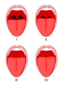
Tonsil Size Scoring

Tonsil Size Grading

  • Grade 0: Tonsils absent
  • Grade 1: hidden behind tonsillar pillars
  • Grade 2: Extend to pillars
  • Grade 3: Visible beyond pillars
  • Grade 4: Enarged to midline


  • Class 1: Tonsil pilllars, Soft palate, and uvula visible
  • Class 2: Tonsil pillars and soft palate visible,  upper portion of uvula
  • Class 3: Base of uvula, soft palate
  • Class 4: Only hard palate


Barbituates – Potent suppressors of REM and may increase spindle activity.

BNZs – potent suppressors of N3 and may increase spindle activity.

MAOIs – more potent that TCA and SSRIs in suppressing REM. MAOIs increase concentrations of Serotonin, NE, and dopamine.

SSRIs – disrupt sleep continuity and suppress REM.

Bupropion – D> NE reuptake inhibitor and also acts as a nictonic Ach receptor antagonist.

Zolpidem – rapidly absorted, T1/2 1.5-2.5 hrs, acts on type 1 GABA-A BNZ receptors. May cause amnesia and parasomnia behavior.

Antipsychotics – Olanzapine, Quetiapine, Ziprasidone decrease SL and improve sleep continuity. REM suppression is variable. Increases in N3 are observed only with Quetiapine. (Seroquel)

Trazodone – inhibits reuptake of serotonin, blocks alpha-1 and histamine (H1) receptors. Half-life 9 hours.

GHB – increases brain dopamine levels and acts on GHB and GABA-B receptors; increases SWS in healthy adults

Drugs which can trigger nightmares – beta-blockers, anti-depressants, barbituates, alcohol, stimulants, steroids

Beta blockers – can decrease melatonin release. May cause sleep disruption, insomnia, nightmares, depression, etc.

Prazosin – alpha 1 antagonist at low dose has been used in PTSD patients resulting in increased TST, REM, and sleep quality and reduced nightmares.

NSAIDs – can disrupt sleep by suppressing normal surge in Melatonin, decreasing prostaglandin D2 synthesis, & attenuateing  the normal nocturnal decrease in body temp.

Drugs MOA Half Life Notes
SARIs(Trazodone)
  • 5HT antagonist, and reuptake inhibitor
  • Block α-1 & H1 receptors.

9 hours

  • Priapism
  • Weight Gain
  • Orthostatic hypotension
  • Arrhythmias in patients with preexisting heart disease
  • ↑TST ↓SL↑ SWS andminimal ↓REM
  • Urinary retention  
NaSSA (Mirtazapine)
  • Central α-antagonist (increasing NE & 5HT)
  •  5HT 2/3 and H1 antagonist

20-40 hours

Weight Gain↑TST↑SWS
TCAs (Doxepin, Amitrptyline, Trimipramine, etc)  
  • 5HT and NE reuptake inhibitor
  • H1 and α-1 antagonist

15-20

  • Doxepin levels increased by cimitedine
  • Exacerbation of narrow angle glaucoma
  • Prolonged QRS
  • Urinary retention  
  • ↓↓REM
  • ↑ PLM
  • ↑ TST


MAOIs
  • ↑ 5HT, D, NE


  • ↓↓↓REM


SNRIs (Venlafaxine)
  • 5HT and NE reuptake inhibitor


  • ↓↓REM
  • ↑ PLM


NDRI (Bupropion)
  • NE &D reuptake inhibitor


  • ↓SWS ↓ PLM
  • ↑REM
  • ↑ Sleep efficiency
  • Nightmares
  • Lowers SEIZURE threshold
BNZs  


  • ↓SWS
Modadifinil  

9-14 hours

 
Armodafinil (R enantiomer)  

15 hours

 
Caffeine
  • Adenosine receptor antagonist

3-5 hours

  • ↓SWS
  • ↑ SL
Dopamine Receptor Agonists  


  • Sudden sleep attacks
  • Compulsive behavior
Medications that can worsen RLS
  • Antipyschotics
  • Antiemetics
  • Antihistamines
  • Anticonvulsants
  • Antidepressants
  • H2 blockers
  • Ca channel blockers


Drugs which can ↑ SWS
  • AEDs (except Lamictal)
  • Anti-psychotics (except Seroquel)
  • Trazodone
  • Mirtazapine
  • GHB


Drugs which can ↑ REM
  • Beta blockers
  • Acetylcholinesterase inhibitors
  • Prazosin
  • THC


Drugs which can ↓REM
  • Steroids
  • MAOIs
  • SSRIs
  • TCAs
  • Venlafaxine
  • Trazodone
  • Opiates
  • H1 antagonists
  • Alcohol
  • Amphetamines, Cocaine


Drugs which can ↓ SWS
  • Caffeine
  • Nicotine
  • Benzodiazepines


 Technology of Sleep Studies

Impedance of EEG & EOG must be < 5kΩ

EOG – referenced to M2

EEG:

  • F4, C4, 02 referenced to M1 [recommended]
  • Fz-Cz, Cz-Oz, C4-M1 is an acceptable alternative deriviation

Digital Accuracy – dependent on digital resolution & sampling rate

  • Digital Resolution (y axis): minimum 12 bit
  • Sampling Rate: dependent on type of signal – the AASM has established standards for low and desired sampling rates

Filters: The AASM has established standards for HFF and LFF filters

Signal

Sampling Rate (Hz)

Filter Settings (Hz)


Minimum

Desired

Low f filter

High f Filter

EEG

200

500

0.3

35

EOG

200

500

0.3

35

EMG

200

500

10

100

ECG

200

500

0.3

70

Snoring

200

500

10

100


Oximetry

10

25


Airflow

25

100

0.1

15

Nasal Pressure

25

100

0.1

15

Rib Cage and Abdomen

25

100

0.1

15


Body Position

1

1


Notes:

For more detailed EEG analysis – increase the sampling rate and HFF settings, but make sure sampling rate is ≥ 3 x HFF setting.

Display Parameters: Minimum 1600 x 1200 resolution or 1950 x 1080

Recognizing Waveforms – defined by amplitude, frequency, waveform morphology, and distribution


Alpha

LAMF

Vertex

Spindle

K complex

Slow Wave

Amplitude

Not defined

Low

Not defined

Not defined

Not defined

75 µV

Frequency

8-13Hz

4-7Hz

≥2Hz

11-16Hz

< 2Hz

0.5-2Hz

Waveform

Sinusoidal

Mixed

Sharp

Distinct

Negative, followed by positive

Not defined

Distribution

Occipital

Not defined

Central

Central

Frontal

Frontal

*Note similarities between K complex and Slow Waves

Sawtooth waves – 2-6Hz, biggest in central channels often observed just before a rapid eye movement.

Eye Movements – downward deflection (indicating positivity) occurs when eye moves in the direction of that sensor. For example, with movement of the eyes to the left, the LEOG will show a downward (positive) inflection, whereas the REOG will show an upward (negative) deflection. In other words – eyes are moving in direction of positivity. With eye blinks, due to bell’s phenomenon, there is a downward (positive) deflection, which is most prominent in the REOG.

Adult Sleep Stage Scoring Rules

W, N1:  see table below.

Stage N2: Score N2 if K complex without arousal or spindle is present in the 1st half of the epoch or was present in the 2nd half of the prior epoch. In the absence of K complexes-without arousal or spindles → continue to score N2 until one of the following events occurs:

  • Transition to W, N3, or R
  • An arousal (first half of the epoch) → score as N1. *Note: EEG arousals include K complexes-with arousal
  • A major body movement (first half of epoch) followed by SEM and LVMF without K complexes/spindles→ score as N1
  • A major body movement (first half of epoch)  occurs with some alpha→ score as W
  • A major body movement occurs (first half of epoch) without alpha and without SEM+LVMF → continue to score as N2

Stage 3: Slow waves (0.5-2Hz) with amplitude > 75µV account for 20% (6s) of the epoch. * Note: Spindles may be observed in Stage 3 sleep

REM: Score R if observe LVMF, low chin tone, and REMs. In absence of REMs, continue to score R if EEG continues to show LVMF and chin tone remains low.  Stop scoring R if one of the following events occurs:

  • Transition to W, N3
  • An increase in chin tone consistent with N1 (first half of epoch) → score as N1
  • An arousal (first half epoch) associated with SEM and LVMF → score as N1. * Note if the arousal is not associated with SEM+LVMF → continue to score as R.
  • A major body movement (first half of epoch) followed by SEM and LVMF without K complexes/spindles→ score as N1
  • A major body movement (first half of epoch)  occurs with some alpha→ score as W
  • A major body movement occurs (first half of epoch) without alpha and without SEM+LVMF and chin tone remains low → continue to score as R.
  • A non-arousal K complex or spindle (first half of epoch) without REMs → score as N2

Scoring epochs between N2 and R

In the absence of spindles and non-arousal K complexes can go back and score prior epochs as R even in the absence of REMS if chin tone remains low.

Adult Sleep Stage Scoring Table


Wake

N1

N2

N3

R

Pts with α

Alpha > 50% of epoch

Alpha < 50% of epoch

K complex without arousal or spindle in first half of epoch or in 2nd half of prior epoch

≥ 20% Slow wave activity

LVMF EEG, low chin tone, REMs

Pts without α
  1. Eye blinks (0.5-2Hz)
  2. Reading eye movements
  3. Irregular eye movements + high chin tone
  •  4-7Hz with slowing
  • Vertex sharp waves
  • Slow eye movements

N/A

N/A

N/A

Definitions:

Arousal – shift to faster EEG for 3 seconds if prior 10 seconds were sleep. If arousal occurs in REM this shift in EEG must be accompanied by an increase in chin tone lasting 1 second.

Major Body Movement – Movement that obscures EEG for more than half of the epoch to the extent that the sleep stage cannot be determined.

Pediatric Sleep Stage Scoring  


Wake

N1

N2

N3

R

Pts with DPR

≥50% epoch has age appropriate DPR over the occipital region

DPR replaced by LVMF for > 50% of epoch

K complex without arousal or spindle in first half of epoch or in 2nd half of prior epoch

(same as adults)

≥ 20% Slow wave activity

(same as adults)

LVMF EEG, low chin tone, REMs

(same as adults)

Pts without DPR
  1. Eye blinks (0.5-2Hz)
  2. Reading eye movements
  3. Irregular eye movements + high chin tone
  • Slowing of background 1-2Hz
  • Slow eye movements
  • Vertex waves
  • Rhythmic anterior theta
  • Hypnagogic hypersynchrony

N/A

N/A

N/A

Pediatric Definitions

Vertex Waves – Broad vertex waves (<0.5 s) can be seen over central regions by 6 months, but vertex waves resembling adult vertex waves usually first appear at 16 months.

Rhythmic anterior theta activity (RAT) – Runs of rhythmic theta activity, maximal over frontocentral regions. May first appear at age 5; common in adolescents and young adults.

Hypnagogic Hypersynchrony (HH) – Paroxysmal bursts of diffuse high amplitude sinusoidal 75-350µV, 3-4.5Hz waves which begin abruptly often maximal over the frontocentral regions. HH often disappears with deeper stages of NREM sleep. HH is seen in 30% at 3 months, 95% of children between 6-8 months, and is less prevalent after age 4-5.

Posterior slow waves of youth (PSW) –  Intermittent runs of bilateral, often asymmetric 2.5-4.5Hz slow waves, which ride upon the dominant posterior dominant rhythm (DPR). The voltage is usually < 120% of DPR voltage. Uncommon in children less than 2, maximal incidence in children 8-14 and uncommon after age 21.

Occipital Sharp Waves with eye blinks – sharp occipital waves < 200 µV usually lasting 200-400 msec which follow an eye blink or eye movement. The initial component of the wave is surface positive.

Development notes:  Drowsiness in Infants up to 8 months of age is characterized by diffuse high amplitude (75-200 µV) 3-5Hz activity.  Drowsiness in children 8 months to 3 years is characterized by runs of high amplitude (75-200 µV) rhythmic/semi-rhythmic  3-5Hz activity often maximal over the occipital regions and/or higher amplitude (>200 µV) 4-6Hz activity over the frontocentral regions. Sleep onset from age 3 is often characterized by 1-2Hz slowing of the DPR or the DPR becomes more diffusely distributed and then replaced by LVMF

Pediatric Sleep Development

EEG finding

Age of Appearance

Trace Discontinue 26-30 weeks
Delta brushes 28-30 weeks, disappear at term
REMs 30 weeks
Trace Alternant 36 weeks, disappears by 12 weeks post-term
Spindles 2 months
K Complex and SWA 4- 6 months
Ability to discriminate N1/2/3 5-6 months
DPR: 2 months 4Hz
6 months 6Hz
3 years 8Hz
9 years 9Hz

Cardiac Rules

  • Sinus Tachycardia during sleep: sustained HR > 90 in adults
  • Sinus Bradycardia during sleep: sustained HR < 40 for ages 6-adult
  • Wide-complex tachycardia: minimum of 3 consecutive beats of a rate > 100/min with QRS ≥ 120msec
  • Narrow-complex tachycardia: minimum of 3 consecutive beats of a rate > 100/min with QRS < 120ms

Movement Scoring Rules

Leg Movement: 0.5 – 10 second duration, amplitude ≥ 8µV above resting EMG. The end of a LM is defined as a period lasting ≥ 0.5 seconds during which the EMG does not exceed 2 µV above resting EMG.

PLM : ≥ 4 LMs separated by a period between 5 – 90 seconds.

  • Leg movements on 2 different legs separated by < 5 seconds count as a single leg movement.
  • LMs occurring 0.5 seconds before/after an anpea or hypopnea are not to be scored.
  • PLM is to be considered associated with arousal if the arousal occurs within 0.5 seconds.
  • Impedence should be < 10,000Ω although < 5,000Ω is preferred.

Alternating Leg Muscle Activation (ALMA) – ≥ 4 bursts of ALMA with frequency of 0.5 and 3Hz. [optional to score]

  • Each ALMA usually lasts 100-500 msec.
  • ALMA may be a benign phenomenon.

Hypnagogic Foot Tremor:  ≥ 4 bursts of activity with frequency of 0.3-4Hz. [optional to score]

  • Each foot movement usually lasts 250-1000 msec
  • May be a benign phenomenon

Excessive Fragmentary Myoclonus: maximum duration 150 msec, must be recorded with at least 20 min of NREM sleep with a frequency of at least 5Hz. Also believed to be benign. [optional to score]

Bruxism : Consists of phasic/tonic elevations of chin tone that are at least twice that of background EMG. Scored if the elevations in tone last 0.25-2 seconds and if three or more occur in sequence. Sustained elevations > 2 seconds if occurring in a sequence of three are also to be scored. Finally, bruxism can be scored by microphone if 2 or more episodes of teeth grinding are heard.

Rhythmic Movement Disorder:  ≥ 4 or more movements with a frequency of 0.5 to 2 Hz. The burst of EMG activity is at least twice that of the background.  

Movement

Rules for Scoring

Leg Movement 0.5 – 10 second duration, amplitude ≥ 8µV above resting EMG
Periodic Limb Movements ≥ 4 LMs separated by a period between 5 – 90 seconds.
ALMA ≥ 4 bursts of ALMA with a frequency between 0.5 and 3Hz
Rhythmic Movement Disorder ≥ 4 movements with a frequency of 0.5 to 2 Hz
Hypnagogic Foot Tremor ≥ 4 bursts of activity with a frequency of 0.3-4Hz.
   
Excessive Fragmentary Myoclonus Maximum duration 150ms, 20 min NREM, 5Hz
Bruxism ≥ 3 consecutive bursts lasting 0.25 – 2 seconds. Or microphone.


Respiratory Scoring Rules

  1. The event duration is measured from the nadir prior to the first breath that is reduced to the beginning of the first normal breath.
  2. If baseline breathing amplitude cannot be determined, events can be terminated when there is an increase in breathing amplitude or where there is resaturation of ≥ 2%

Adult Respiratory Event

Scoring Rules


Apnea [recommended]
  1. Drop in thermal sensor by ≥ 90% for 90% of event duration
  2. Duration ≥ 10 seconds
Hypopnea   [recommended]
  1. Drop in nasal pressure by ≥ 30% for 90% of event’s duration
  2. Duration ≥ 10 seconds
  3. ≥ 4% desaturation
  1. Hypopnea [alternative]
  1. Drop in nasal pressure by ≥ 50% for 90% of event’s duration
  2. Duration ≥ 10 seconds
  3. ≥ 3% desaturation
RERA [option]

Flattening of nasal pressure for ≥ 10 seconds or increased respiratory effort leading to arousal, not fulfilling criteria for apnea/hypopnea.

Cheyne Stokes Respiration

≥ 3 cycles of crescendo-decrescendo breathing and at least one of the following:

  1. ≥ 5 central apneas per hour
  2.  Cyclic crescendo-decrescendo breathing for ≥ 10 consecutive minutes
Hypoventilation ≥ 10mmg increase in PCO2 during sleep as compared to wake supine value


Pediatric Respiratory Event

Scoring Rules


Obstructive Apnea
  1. Obstructive event lasts ≥ 2 breaths determined by baseline breathing pattern
  2. Drop in thermal sensor by ≥ 90%  for 90% of event’s duration
  3. Event associated with continued respiratory effort
Central Apnea
  1. The event lasts  ≥ 20 seconds and at least 2 breaths
  2. Associated with either arousal/awakening or ≥ 3% desaturation.
  3. Absence of respiratory efforts
Mixed Apnea
  1. Event lasts ≥ 2 breaths determined by baseline breathing pattern
  2. Drop in thermal sensor by ≥ 90%  for 90% of event’s duration
  3. Absent effort initially followed by resumption of effort prior to end of event.
Hypopnea
  1. Drop in nasal pressure by ≥ 50% for 90% of event’s duration
  2. Event lasts ≥ 2 breaths
  3. Event is associated with arousal/awakening/or ≥ 3% desaturation.
RER
  1. Drop in nasal pressure by < 50%
  2. Event lasts ≥ 2 breaths
  3. Event accompanied by snoring, noisy breathing, elevation in pCO2 or visual evidence of increased work of breathing

Or when using esophageal monitoring:

  1. Progressive increase in respiratory effort
  2. Event lasts ≥ 2 breaths
  3. Event accompanied by snoring, noisy breathing, elevation in pCO2 or visual evidence of increased work of breathing
Periodic Breathing > 3 episodes of central apnea lasting > 3 sec separated by ≤ 20 sec of normal breathing
Hypoventilation When > 25% of TST is spent with PCO2 > 50  
In pediatrics, the thermal sensor can be used for scoring hypopneas if necessary, but, only a nasal transducer or esophageal monitor can be used for scoring RERAs.